技術開発・イメージング
RNA Imaging & Technology Development
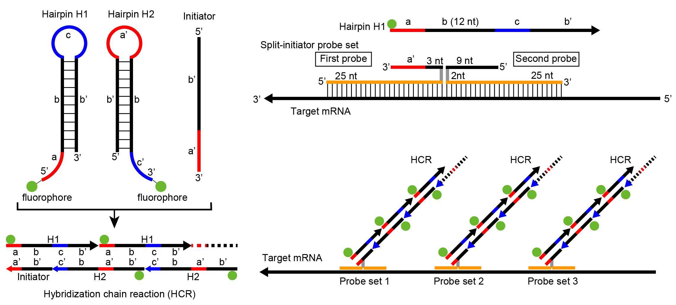
Principle of isHCR for sensitive and multiplex RNA visualization
多様な細胞から構成される脳の機能と構造を理解するためには、個々の細胞におけるRNA発現を高感度かつ多重に可視化する技術が不可欠です。従来のin situ hybridization法は、標識プローブと標的mRNAの相補的結合を利用しますが、感度や特異性、多重染色に要する時間、Proteinase K処理による組織障害や抗原性の低下などの課題があります。近年、新しいRNAイメージング法も開発されていますが、多くは高価な試薬や専用装置を必要とし、広く普及するには課題が残されています。
恒岡准教授を中心に、ネッパジーン株式会社との共同研究により、 isHCR法を基盤に高感度・低ノイズ・低コストなRNAイメージング技術を開発しました。複数のRNAを簡便かつ高速に同時検出できるうえ、Proteinase K処理を必要としないため免疫染色との併用が可能です。また、組織の自家蛍光を効率よく消光する TiYO™ も独自に開発・製品化されました。これらの技術は特許化され、ISHpalette™ および TiYO™ として製品化されています。
ISHpalette™ および TiYO™ を組み合わせることで、組織の自家蛍光を大幅に低減しながら、高感度・低ノイズでRNAシグナルを検出できるため、従来は困難であった組織や生物種にも応用できるようになりました。哺乳類に加えて魚類、鳥類、両生類、爬虫類、昆虫など多様な生物種の組織へ応用が広がり、神経科学のみならず発生学、比較生物学、再生生物学など幅広い生命科学分野で利用されています。
The brain is composed of hundreds of cell types, each with a distinct gene expression profile. visualize RNA expression in individual cells with high sensitivity and multiplex capability. Conventional in situ hybridization relies on complementary binding between labeled probes and target mRNAs, but it has several limitations, including insufficient sensitivity and specificity, and loss of antigenicity caused by Proteinase K treatment. Although newer RNA imaging methods have improved sensitivity and reduced background, many require expensive reagents or specialized equipment, limiting their widespread use.
Led by Associate Professor Tsuneoka, in collaboration with Nepa Gene Co., Ltd., we developed a highly sensitive, low-noise, and cost-effective RNA imaging technology based on in situ Hybridization Chain Reaction (isHCR). Our method preserves tissue morphology and antigenicity without requiring Proteinase K treatment, enabling seamless combination with immunohistochemistry. It also allows multiple RNA species to be detected rapidly and simultaneously using a simple workflow.
In parallel, we developed TiYO™, a proprietary technology that efficiently quenches tissue autofluorescence. By combining ISHpalette™ with TiYO™, tissue autofluorescence can be markedly reduced while maintaining highly sensitive, low-background RNA detection. This combination has substantially expanded the range of tissues and organisms that can be analyzed. These technologies are now being applied to tissues from a wide variety of organisms, and are contributing to diverse fields such as developmental biology, comparative biology, regenerative biology, and many other areas of life science.
Selected publications
Tsuneoka et al., Front Mol Neurosci 2022
Tsuneoka et al., Front Mol Neurosci 2020